Pioneering Innovations in Medical Imaging for the Central Nervous System (CNS)

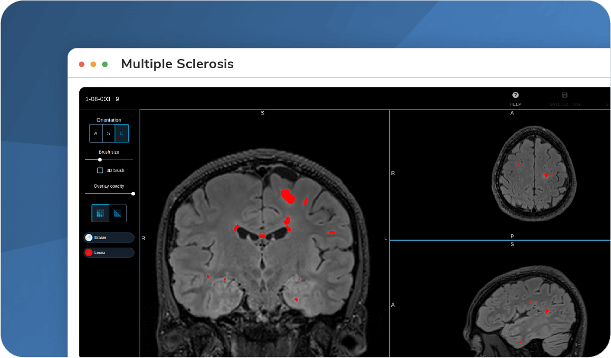
Multiple Sclerosis
State-of-the-art imaging biomarkers and tools for lesion segmentation, activity, load, diffusion, and volume.
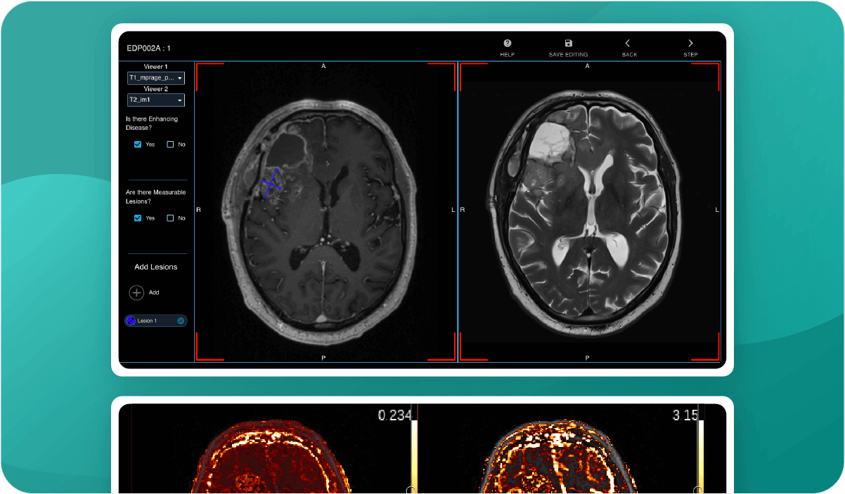
Neuro-Oncology
AI high-tech tools and imaging biomarkers for brain oncology, RANO assessment, contrast, perfusion, tumor volume, and Gadolinium.
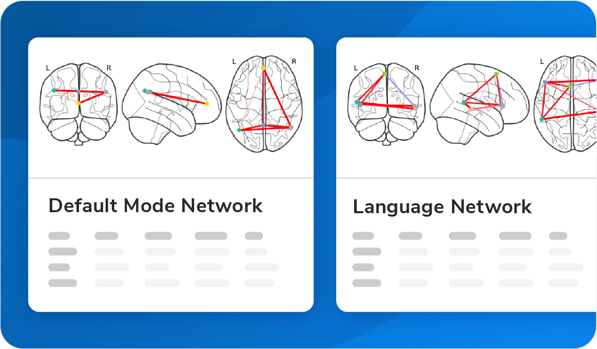
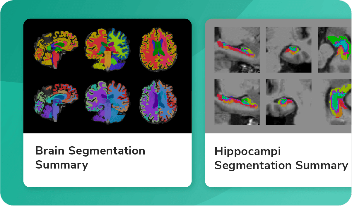
Neuropsychiatric Diseases
Cutting-edge imaging biomarkers for connectivity, volume, and thickness.
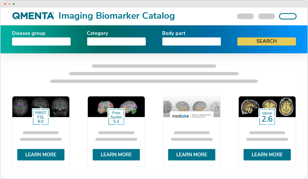
Visit our Imaging Biomarkers Marketplace