The Artificial Intelligence (AI) boom is more than just a fleeting trend. Did you know that AI is now capable of analyzing complex medical images with accuracy surpassing human experts? Almost every day, headlines showcase breakthroughs from companies like OpenAI, Anthropic, Google, or IBM. Everything indicates that AI is here to stay. Its fields of application are huge, including the analysis of medical images. It is currently being applied to detect different types of cancer (brain, breast, lung, skin, colon, …), Alzheimer's, stroke, and COVID-19 among others.¹
In this post, we explore how AI technologies not only provide benefits but can also seamlessly integrate into medical imaging workflows, offering unprecedented efficiency and accuracy.
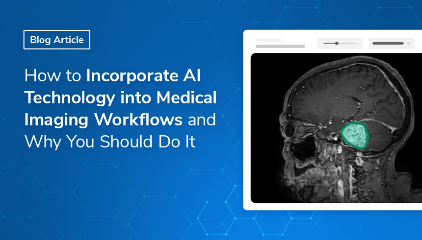
How AI Integrates into a Medical Imaging Workflow: A Practical Example
A medical imaging workflow is the sequence of steps necessary for the imaging procedure, from acquisition and storage to interpretation and sharing. Technology-supported medical imaging workflows are becoming a key component in medicine, including research, clinical trials, and routine clinical practice. These workflows are created with the potential to reduce the amount of workload and cost while improving data management, collaboration, and diagnosis. Integrating AI into a medical imaging workflow can boost productivity even further.
Let’s imagine a clinical trial in which the safety or efficacy of a new drug is tested for the treatment of brain tumors, with several sites participating in the study. In this scenario, some kind of medical imaging technique will most likely be needed for in vivo quantification of the effect of the drug. This means a substantial quantity of data influx at various points of the study.
AI can be of great benefit in many steps of the processes involved. However, the greatest direct impact can be achieved by employing AI algorithms that will assess the imaging data in a consistent and seamless way. The added value that AI can bring is enormous and will be discussed later. Having all steps of the data acquisition and management of all study sites centralized, preferably in the cloud, opens up possibilities to add AI at various steps of the flow.
How simple it can be to integrate AI into existing medical imaging workflows? Before addressing this question, let's first examine some benefits and current challenges of AI technologies.
Five Benefits of AI in Medical Imaging
This iterative hybrid process can be integrated into any of the QMENTA tools in a very simple way.
Why do we want to integrate AI into medical imaging workflows? The straight answer is that AI can be better than humans in certain tasks, and thus can help healthcare professionals in their diagnosis and treatment decisions. Let’s explore this in more detail.
Reproducibility
An AI algorithm, if well done, will always provide the same (or at least similar) results after analyzing the same image. This is something that humans cannot do. A medical imaging expert may assess the same image differently depending on his/her state of mind. Several factors may affect their objectiveness such as fatigue, time pressure, or even mood. Moreover, the same medical image may be assessed differently by different professionals. AI algorithms clearly have more reproducible outcomes in that sense.
Minimizing human errors
Because AI algorithms are made by humans, they will most likely include design errors. However if they are properly designed, the error will remain constant. And as explained above, they will show a reproducibility that humans do not have.
Another advantage is that an AI algorithm can improve with time and the intrinsic human error can be minimized. With proper training, the AI will get more robust and precise as it analyzes new images.
Recognizing complex patterns.
AI is better at analyzing medical images than humans. They can provide new insights about what features should be valued to support decisions, helping healthcare professionals to better predict disease outcomes.
Early-stage disease detection
When analyzing medical images, the speed and precision of AI systems can help in identifying diseases at an earlier stage than a human could. Certain patterns/lesions might not be visible to a human but are identifiable to an AI algorithm. This is crucial as detecting a disease as early as possible can not only improve treatment outcomes but also save lives.
Time and cost-effectiveness
Computers are much faster than humans in many tasks, including assessing complex medical images. Where a human can take hours, an AI can seconds. As an example of such fast algorithms, FastSurfer² can segment a whole brain into 95 substructures in less than a minute.
Key Challenges When Deploying AI Tools in Medical Imaging
While the advantages can be significant, there are also some challenges regarding AI technologies worth discussing. For instance, AI models often require very large data sets for training, and it is neither easy nor cheap to gather nor easy to access them. On the one hand, it is not a simple task to organize and pre-process data generated from different institutions, and on the other, image-data sharing is very limited due to HIPAA regulations.
Furthermore, AI models can suffer from “hallucinations”³ which can lead to incorrect results. This can be due to different factors such as insufficient or biased training data, or incorrect assumptions made by the model.
Another challenge is related to the manipulation of medical data. This is a very sensitive matter and there are strict regulations and compliance issues. It is not the same to have AI generate a picture or a text for recreational purposes as compared to applying AI for treating sensitive and oftentimes critical medical data. Taking this into consideration, ensuring the security of the data is paramount, be it in a local or cloud deployment.
Finally, integrating AI tools into existing medical imaging workflows might look extremely complicated, and this thought might be discouraging. But, with the proper approach, this is far from reality.
The QMENTA Approach: Integrating AI Tools in 3 Steps
- The QMENTA Imaging Hub is a cloud-based computing platform for securely uploading, storing, and processing medical images. Within its main advantages, you can find:
- No need to install anything on your local computer. You just need an internet connection and a web browser.
- Our AI based data uploader tool recognizes the different medical images types and modalities and allows you to keep your data structures and harmonized.
- The uploader tool also strips all PHI information from the medical images and is HIPAA-compliant.
- Being cloud-based, it makes collaboration between different sites very easy, no matter the physical distance.
- Since it is cloud-based, we have access to unlimited memory storage for our medical data, which usually is quite heavy.
- Access to a large existing tool catalog for analyzing your different medical images.
- Again, since it is cloud-based, access to unlimited computing power to run the different tools. And also the possibility to run tools in parallel, saving time.
- Our SDK allows you to easily create and integrate new AI solutions to existing medical imaging workflows.
This last point is the one we are going to explain in better detail in the rest of this section.
The deployment of the AI tool consists of 3 steps:
- Write the main program (or use an existing one).
- Build and push a Docker container with the appropriate environment for the tool to run.
- Add the tool to the QMENTA Imaging Hub.
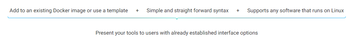
You can start by writing and testing your AI tool in your local environment. Any programming language can be used since QMENTA works with containerized images, and this step can easily be done with Docker.
Finally, you will need developer access to the QMENTA I, which allows you to create and manage new tools. This includes the ability to define how the tool is presented and configured. Then it can be made available in the QMENTA tools catalog for other users. You can find more detailed info in the SDK documentation.
Note that it is possible to deploy already existing AI tools instead of writing your own code. In fact QMENTA already offers different third-party tools such as FreeSurfer, AFNI, ANTs, FSL, RADIOMICS, MRtrix3, etc.
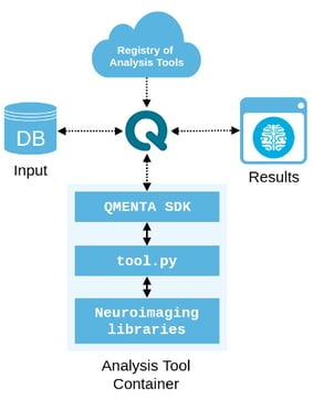
A visual representation of the QMENTA SDK and how it integrates into the workflow
The main advantages of using the QMENTA SDK are that it allows researchers to use Docker images with pre-installed imaging development environments which:
- Eliminate tedious virtual machine setup and the need to build/maintain a local cluster.
- Combine public/open resources: NeuroDebian, Jupyter Notebooks, and QMENTA API as desired.
- Integrate data warehousing and processing with QMENTA API.
This approach allows you to:
Develop and Test
Easily develop, test, and run tools in a scalable cloud environment. Run Docker containers with custom tools in fully portable environments.
Share
Quickly share and benchmark your tools and workflows with the medical imaging and neuroimaging community.
Market
Distribute and monetize your research with a simple revenue-sharing model. Bridge the gap between your innovation and clinical practice.
Tool creation with the QMENTA Imaging Hub
Why Cloud-Based AI is the Future of Medical Imaging Workflows
As we have seen, the benefits of cloud-based AI are enormous, and its applications are revolutionizing many areas, including the medical realm. Of course, there are some challenges to overcome, but with a proper approach, we can easily profit from its use.
Nowadays, medical imaging workflows are widely used in medical applications, and if its design is modular, the integration of AI tools should be quite straightforward. This is the case with the QMENTA Imaging Hub, an all-in-one place to keep your data and your analysis tools.
As it is cloud-based based you don’t need to install anything into your local computer and that allows for seamless integration of your AI tool into your medical imaging workflow. This cloud approach means that you would have access to virtually unlimited data storage and unlimited computational resources.
Once you have integrated your AI tool, you will not only save time and money, but you will get more reliable results in your medical image assessments. Our calculations show that by using advanced image analysis you can reduce time by 80% and cost by 90%.
Finally, the QMENTA Imaging Hub is very well suited for collaboration between sites and moreover it allows you to share and market your own developed AI algorithms so the rest of the medical imaging community can benefit from them. And even more, beyond saving money, you can even make a profit!
Frequently Asked Questions
What are the main benefits of integrating AI into medical imaging workflows?
AI integration in medical imaging offers five primary advantages: reproducibility — AI algorithms produce consistent results regardless of fatigue or subjective state, unlike human reviewers; reduced human error — well-designed AI algorithms maintain a constant, predictable error profile; pattern recognition — AI can identify complex features in imaging data that exceed human visual detection; early-stage disease detection — AI can detect subtle patterns indicating disease before they are visible to a human reader; and time and cost efficiency — AI systems can process images in seconds compared to hours for manual analysis, with some tools like FastSurfer completing full brain segmentation in under one minute.
What challenges should teams expect when deploying AI tools in medical imaging?
The main challenges are: data requirements — AI models require large, well-organised training datasets that are difficult and expensive to assemble, particularly given HIPAA restrictions on image sharing; AI hallucinations — AI models can produce incorrect results due to insufficient or biased training data, making validation essential before clinical or trial use; data security — medical imaging data is highly sensitive, requiring strict regulatory compliance for both local and cloud deployments; and workflow integration — while technically straightforward with the right infrastructure, integrating AI into existing systems requires planning and the right development environment.
How does QMENTA enable AI tool integration in medical imaging?
QMENTA's Imaging Hub is a cloud-based platform for securely uploading, storing, and processing medical images, with access to unlimited storage and computing power for running AI tools. Integration of a new AI tool follows three steps: writing or adapting the main AI algorithm; building and pushing a Docker container with the appropriate environment; and adding the tool to the QMENTA Imaging Hub using developer access. The platform's SDK supports any programming language through containerised images, and tools can be distributed to the broader QMENTA research community through the tools catalogue, including through a revenue-sharing model.
What is an AI hallucination in medical imaging and how can it be mitigated?
An AI hallucination in medical imaging refers to a situation where the model produces results that appear plausible but are incorrect — for example, incorrectly segmenting a region as a lesion or misidentifying tissue type. This occurs due to insufficient training data, biased datasets, or incorrect model assumptions. Mitigation strategies include training on large, diverse, and well-annotated datasets; external validation on data from different sites and scanners; prospective clinical validation rather than solely retrospective testing; and human-in-the-loop review workflows where AI outputs are reviewed and refined by qualified experts before clinical or regulatory use.
References:
¹ Abdulhamit Subasi, ed., Applications of Artificial Intelligence in Medical Imaging, Artificial Intelligence Applications in Healthcare and Medicine (London San Diego, CA Cambridge, MA Kidlington, Oxford: Academic Press, an imprint of Elsevier, 2023).
² Henschel et al. FastSurfer - A fast and accurate deep learning-based neuroimaging pipeline. NeuroImage, 2020
³ IBM, “What Are AI Hallucinations?“, 11. April 2024, https://www.ibm.com/topics/ai-hallucinations.