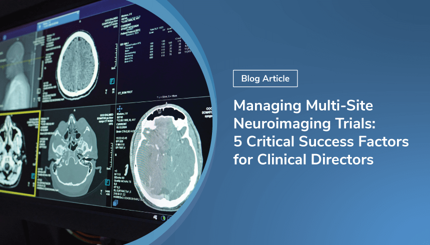
Reflections from RSNA 2025: AI is everywhere, but are we solving the right problems?
RSNA 2025 highlighted a crowded AI landscape—but also the growing complexity of multi-site imaging trials. Learn why scanner variability, protocol...
Learn the 5 critical success factors clinical directors need to run multi-site neuroimaging trials—covering protocol standardization, QC, training, and scalable technology.
Multi-site neuroimaging trials terrify most first-time clinical directors, and for good reason. You're coordinating imaging across facilities running different scanner manufacturers, software versions, and quality control philosophies. Protocol deviations multiply. Sites drop out mid-trial. Suddenly you're staring at a dataset with 30% unusable scans and a looming regulatory deadline.
Yet we keep doing multi-site trials because we must. Single-center studies can't enroll fast enough or generate the statistical power needed for modern regulatory standards. You need those 200+ patients, which means multiple sites, which means imaging standardization that actually works. The budget hit is real (expect 30-50% increases from imaging CRO fees, training, quality control infrastructure, and data management), but the difference between success and failure usually comes down to five critical factors.
Most directors hit the same problem early: you need detailed imaging protocols to assess whether sites can deliver, but you need site input to develop protocols that won't cause rebellion three months into enrollment. The solution is to iterate collaboratively.
Your charter is the trial's imaging constitution, the document that sites, regulators, auditors, and your Data Monitoring Committee will reference when questions arise. It covers the entire lifecycle: acquisition standards, quality control procedures, reader certification, data handling specifications, and regulatory compliance documentation. For FDA-regulated trials, you're aligning with their "Standards for Clinical Trial Imaging Endpoints" guidance and demonstrating 21 CFR Part 11 compliance.
Charter development isn't just documentation—it's a significant investment that prevents downstream failures. Industry data shows that site start-up costs can run over $30,000 with charter development consuming a substantial portion. Budget 8-12% of your total imaging budget ($32-48K for a 200-patient trial at $2K per scan) upfront for charter development, site qualification, and training.
The charter development process typically spans 4-6 months for standard structural MRI protocols (8-12 months for complex multi-modal studies). However, site activation adds significant time: median activation times are 9.4 months for academic medical centers versus 4.8 months for independent sites—a critical planning factor.
Each day of Phase III trial delay costs approximately $55,716 in direct expenses, meaning every month of start-up delay costs over $1.5 million. More concerning, nearly 43% of total trial expenditures lie in poorly-tracked startup activities, including site overhead and unallocated costs.
Start with vendor-agnostic acquisition parameters with translation tables. When you specify TR of 2000ms, you need documented equivalents for Siemens, GE, and Philips scanners because each vendor interprets timing differently. Quality control thresholds need visual examples—"minimal motion artifact" is meaningless without reference images.
Your data transfer protocols must specify DICOM anonymization standards (HIPAA Safe Harbor compliance), secure cloud transfer with encryption standards, and long-term archiving SOPs meeting regulatory retention requirements.
Don't overlook your incidental findings protocol: Who reviews scans for clinically significant findings? What's reportable? What's the notification pathway? Who pays for follow-up? Get this IRB-approved at each site before enrollment starts.
Map your entire site activation process before starting. Research shows sites can achieve 45.6% reductions in start-up cycle time (from ~25 weeks to ~14 weeks) through process standardization and eliminating unnecessary delays where critical documents sit untouched.
Perfect standardization will kill your trial. Directors who demand uniformity across every parameter end up with site compliance nightmares. Sites drop out. Enrollment slows. You need to be rigid where it matters and flexible where it doesn't.
Think of your parameters in three tiers. Tier 1 parameters are non-negotiable because they directly and significantly impact quantitative biomarkers: sequence type, spatial resolution, field strength, slice thickness. Variation here introduces systematic bias that no post-processing can fully correct. Document in your Statistical Analysis Plan how these parameters relate to your endpoints. Tier 2 parameters are relatively fixed at each site but may vary between sites—coil configurations, parallel imaging acceleration factors, and certain reconstruction algorithms are determined by local hardware and software. The goal here is harmonization: tools like ComBat can retrospectively correct site effects while preserving biological differences. Document these variations in your charter and specify your harmonization approach for regulators. Tier 3 parameters are site discretion items like patient comfort protocols, scheduling preferences, local safety procedures, and scanner booking workflows. These affect operations, not science, so grant sites autonomy here."
Monthly phantom scans catch scanner drift before it corrupts longitudinal data and provide objective evidence of hardware stability. Require scanner qualification before any patient enrolls: back-to-back test-retest scans on three volunteers documenting reproducibility with coefficient of variation below 5% for volumetric measurements.
Here's an uncomfortable truth about software versions: truly locking them for 3-5 year trials is often impossible. Scanner manufacturers push mandatory security updates. Hospitals prioritize patient safety over research protocol adherence. Your charter needs a pragmatic software management plan that requires 30-day advance notice of any software updates, conducts immediate post-update phantom testing, performs statistical comparisons of pre/post-update data, documents all changes in your trial master file, and considers treating post-update data as a separate "site" in harmonization analyses if significant differences emerge. Budget $200-400 per phantom session per site, small insurance against data loss.
Make sure to document your harmonization strategy in detail. Include the specific harmonization algorithms you'll use (ComBat, ComBat-GAM, etc.), validation data demonstrating effectiveness, sensitivity analyses showing results are robust to harmonization approach, and contingency plans if harmonization proves inadequate.
Discovering motion artifacts six months post-scan is like finding foundation cracks after you've built the house. The patient's gone. The rescan window closed. Your sample size just decreased. Real-time quality control transforms this from inevitable to preventable.
Industry best practice calls for centralized quality control review within 48 hours of acquisition. This keeps patients available for immediate rescans and demonstrates to regulators that you're actively monitoring data quality. Implement a three-phase QC architecture to make this work.
In Phase 1, Traditionally, technologists performed visual checks using protocol checklists immediately after scanning—a labor-intensive process prone to human error and inconsistency. Modern imaging platforms have transformed this through automated protocol adherence checking that verifies sequence parameters, anatomical coverage, slice orientation, and file integrity against your imaging charter specifications.
This automation catches protocol deviations before the patient leaves the scanner. Rather than relying on technologists to manually cross-reference dozens of parameters, automated systems flag deviations in real-time, allowing immediate correction while the patient is still on-site.
In Phase 2 certified readers apply standardized rubrics from your imaging charter, catching subtle issues that automated systems miss—borderline motion artifacts, anatomical anomalies, or image quality concerns requiring expert judgment. Your imaging CRO or core lab should document reader credentials, review timestamps, pass/fail determinations, and electronic signatures meeting 21 CFR Part 11 requirements.Phase 3 runs concurrently with Phase 2 and uses automated tools to flag objective metrics falling outside acceptable ranges: signal-to-noise ratio thresholds, motion parameters, geometric distortion indices, contrast-to-noise ratios.
Your charter must specify clear rescan criteria. Mandate rescans for incomplete anatomical coverage, severe motion (greater than 3mm translation), or protocol deviation affecting primary endpoint. Make rescans optional for moderate motion (1-3mm) or minor artifacts not affecting regions of interest. Accept with notation cases of mild motion (less than 1mm) or artifacts outside regions of interest.
Budget $150-300 per scan for central review services—$30-60K for a 200-patient trial. This sounds expensive until you consider discovering 30% unusable scans after database lock.
The combination of automated protocol compliance checking with expert human review creates a robust safety net. Automated systems catch technical deviations immediately, while expert reviewers provide clinical judgment automated tools cannot replicate, dramatically reducing risk of discovering systematic imaging problems late in your trial.
Most site training is theater. A webinar, some slides, maybe a test scan, then everyone signs off and hopes for the best. This approach guarantees protocol deviations.
Nobody talks about the real training gap. MRI technologists are experts at clinical scanning but not experts at research protocols with strict standardization requirements that may conflict with their clinical training. When your protocol says "do not adjust parameters for patient comfort," but their entire clinical career has trained them to optimize image quality through parameter adjustment, you're asking them to override deeply ingrained professional instincts.
Set up competency-based training in three stages, recognizing that sites have limited availability due to clinical scan schedules.
If your trial uses centralized readers for outcome assessment, you need standardized training materials including representative images spanning the range of expected pathology and clear decision criteria for each outcome measure. Your formal certification process should require readers to score a standardized test set with known ground truth, with pass thresholds typically at 80-90% agreement with gold standard. Conduct inter-rater reliability assessment by having multiple readers score the same cases to document consistency. Calculate and report kappa statistics demonstrating acceptable agreement (typically κ greater than 0.7 for primary outcomes).
Training isn't one-and-done. Schedule quarterly calibration sessions where readers score common cases and discuss discrepancies. This prevents reader drift over multi-year trials. Budget $5-10K per site for initial training and $2-3K annually for ongoing calibration.
Your technology stack makes or breaks operational efficiency. The right tools transform chaos into manageable workflows. The wrong ones create bottlenecks that slow enrollment and frustrate sites.
You need automated DICOM reception and routing systems that receive scans from sites, verify protocol compliance, route to appropriate reviewers, and flag deviations in real-time. Manual email-based transfers don't scale beyond 3-4 sites. Cloud-based PACS systems provide the centralized data repository you need, with secure storage, version control, and access management. Look for platforms offering HIPAA-compliant encryption at rest and in transit, role-based access control, audit trails meeting 21 CFR Part 11 requirements, and integration with EDC systems for seamless data flow.
Your quality control workflow management platform should support multi-phase review workflows with automated notifications, standardized scoring forms, and electronic signatures. Readers need side-by-side comparison tools for longitudinal assessments. Modern platforms integrate statistical harmonization tools, automated quality metrics calculation, and real-time dashboards showing enrollment progress, quality trends, and site performance.
QMENTA's imaging platform addresses these requirements comprehensively. Their cloud-based system handles DICOM reception, automated quality control workflows, centralized reading, and statistical harmonization within a single integrated environment. The automated protocol compliance checking flags deviations before human review, while the potential for harmonization algorithms including ComBat and site-effect correction work seamlessly with configurable QC workflows supporting your specific three-phase review process. Their advanced analytics dashboards provide real-time visibility into trial health, and the regulatory-ready documentation includes complete audit trails and electronic signatures.
For multi-site neuroimaging trials, integrated platforms like QMENTA eliminate the inefficiency of stitching together multiple point solutions. Sites upload to one system. Reviewers access one interface. Data flows to your EDC. Harmonization happens within the same environment where data is stored and reviewed.
Some academic centers attempt building custom solutions, but this rarely succeeds for multi-site trials. The regulatory requirements, security standards, and workflow complexity require specialized expertise that most IT departments lack. Budget $50-150K annually for enterprise imaging platforms on 200-patient trials. This includes data storage, QC workflow management, reader access, and technical support. Compare this to the cost of building and maintaining custom infrastructure, which typically exceeds $200K in development costs alone, plus ongoing maintenance.
RSNA 2025 highlighted a crowded AI landscape—but also the growing complexity of multi-site imaging trials. Learn why scanner variability, protocol...
Imaging compliance failures often surface only during FDA inspections. Learn how documentation gaps, audit trails, and delayed QC put trials at risk.
Since the discovery of X-Rays in 1895, Radiology has been evolving rapidly. With 127 years of advancements in mind, what can we expect for its future?