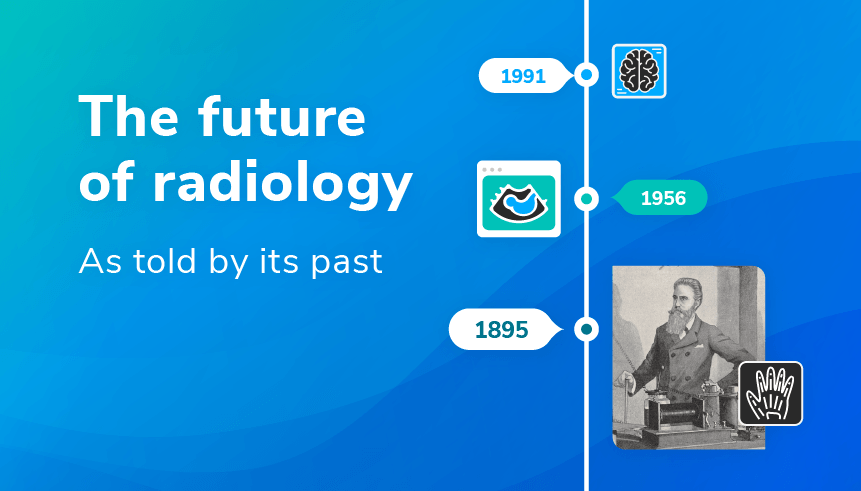
From X-Ray Hysteria to AI Hysteria: A Familiar Pattern in Radiology
“A new scientific truth does not triumph by convincing its opponents and making them see the light, but rather because its opponents eventually die, and a new generation grows up that is familiar with it.”
Planck’s Principle. An argument for why new scientific theories and methods are often slow to be accepted. Unlike the sluggish adoption of heliocentrism, Darwinian evolution, and continental drift theory, radiology appears to have rarely followed suit. The field of radiology was born out of a paradigm shift: the discovery of the X-ray. It took very little for X-rays to capture mass scientific and societal interest, in part due to their undeniable practical applications.
It set the tone for radiology as a rapidly evolving scientific discipline. So with this and 127 years of advancements in mind, what can we expect for its future?
When, in December 1895, Planck’s contemporary and Father of Radiology, Wilhelm Röntgen, handed over his findings to his secretary, it is hard to believe he could have predicted the full extent of their impact. But Röntgen’s famous words, “now, all hell can break loose”, were prophetic after all. X-rays were a scientific success: 1,044 papers on the topic were published in 1896, from physics and medicine to microbiology and food. They were also a commercial success.
Led by Thomas Edison, public exhibitions were held where attendees could do the unthinkable and see into their bodies. Their ability to reveal the invisible meant X-rays found their way into art, comic books, and even shoe shops. However, this quality was unsettling for some. To name the rays after the unknown quantity ‘X’ perfectly captured the sense of intrigue and fear surrounding them, despite their rapid assimilation into society.
There are parallels between the public awe with which X-rays were initially received and society’s collective apprehension towards artificial intelligence (AI). AI Critics have warned of a similar ‘hellish’ future. When we think of how AI is represented in popular culture, we think of highly advanced robots and a technological singularity. And it’s not total misinformation.
In his book The Precipice: Existential Risk and the Future of Humanity, philosopher Toby Ord contemplates a future in which “an ever-increasing share of power is handed over to AI systems” and humans eventually cede control over our destiny. A realistic early step in this would be mass unemployment, and the concern is shared by some radiologists.
However, hypothetical ethical challenges should not overshadow AI’s huge potential. When the British Institute of Radiology (BIR) quizzed presidents of radiological societies from around the world on the future of imaging in 2019, AI was repeatedly touted as a positive development for the field.
AI should not be seen as a replacement for the perceptual expertise of examining a patient scan and making a diagnosis, but rather as an aid to that process. It can assist with the problem of Big Data in the neurosciences and beyond. It can substantially accelerate imaging efficiency, as with deep learning-based image reconstruction technologies. Ultimately, it will allow time spent with the patient to be more fruitful.
The most pressing issue for radiologists in this respect is the use of unrepresentative, low-quality datasets in developing these technologies.
Radiology is the very quintessence of technology’s transformative effect on medical practice. Few fields are better placed to benefit from it, and so, as in the past, new developments should be embraced in the face of public trepidation.
The CT Revolution: How Multidisciplinary Science Transformed Radiology
Computed tomography (CT) was another technological triumph in the history of radiology. Although radiography equipment underwent steady improvements in the early 20th century, X-rays remained poor at imaging soft tissues such as the vascular system and the brain. Contrast media (e.g. barium sulfate) were introduced as a solution to this, but were not without (potentially life-threatening) side effects. X-ray research seemed to have hit a wall.
This all changed at the 1972 Congress of the BIR following Sir Godfrey Hounsfield and Dr. James Ambrose’s presentation: ‘Computerized Axial Tomography’. Using their prototype scanner - a rotating X-ray source, sensors, and computer-, they reconstructed for the first time an image of a brain tumor in the frontal lobe without a contrast agent. Being computer-based rather than film-based, CT enabled clinicians to visualize tissues of interest at a greater magnification and through various planes.
It has since proved indispensable in the clinic for everything from image-guided surgery to radiotherapy planning.
The fascinating thing about CT is that Hounsfield was not the first to conceptualize it. It was envisioned in one of the very first articles to break the news of X-rays to the world in January 1896. The Frankfurter Zeitung spoke of “perfecting the method” so that “only parts of the soft tissue remain transparent” to “expose a deeper slice”. This was not your average newspaper, as it was later one of the few media outlets to not be completely controlled by the Ministry of Propaganda in Nazi Germany.
Then, in 1922, a patent application was filed for the process of tomography by Bocage. His invention involved synchronous movement of the X-ray tube and plate, enabling the blurring of structures above and below a section of interest. Even Hounsfield himself - an electronics and radar man at heart with significant contributions in the computer field - did not originally conceive of the idea with medicine in mind.
He was instead investigating pattern recognition, working under the hypothesis that with sufficient measurements of the exterior of a box, you should be able to work out what’s inside.
What can we take away from this for the future? It teaches us that the breakthrough technologies of the future, or at least the seed of an idea, may already exist. That the boundaries between scientific disciplines are where the magic happens. Radiologists must be equipped to nurture a widening skill-set and be cross-disciplinary thinkers, able to understand the patient as a clinician and the machines as an engineer.
They have to not only learn but also innovate, and at the very least, effectively collaborate.
Whether this is tangible is a different question. Radiology departments across the world are battling a lack of resources. In developed countries, this reveals itself as a shortage of radiologists. In economically developing countries or those with extensive rural populations, unequal access to imaging technologies is a challenge, much less the latest advances. It may be that additional interdisciplinary training is not exactly at the top of many departments’ priorities.
Quantitative MRI (qMRI): Radiology's Shift from Subjective Art to Objective Science
In the BIR’s society survey, the Radiological Society of South Africa had an interesting take: “Radiology will transform from a subjective perceptual skill into an objective science.” Radiology has been moving on this path for some time, but has started to speed up with the advent of magnetic resonance imaging (MRI), in particular the emerging field of quantitative MRI (qMRI).
qMRI goes beyond visual detection of macroscopic tissue pathology and considers intrinsic tissue properties such as longitudinal and transverse relaxation times, susceptibility, axon myelination, and iron deposition. There is a great deal of research going into the development of MRI biomarkers that are both sensitive and specific to certain pathologies.
Researchers are working towards complete characterization of abnormal tissues using, for example, probability maps constructed from both imaging and non-imaging information. This would circumvent the ambiguity and human error that come with manual data reviews and, ideally, the need for biopsies when making a definitive diagnosis. It comes leaps and bounds from the qualitative interpretations of the very first X-ray photographs.
Despite the abundance of quantitative tools in active research, their integration in the clinic has a long way to go. Currently, to truly extract the most from all the information MRI has to offer, data acquisition could take hours. There is also the challenge of analyzing potentially vast quantities of data in a hospital setting. Even then, the MRI community is plagued by reproducibility and repeatability issues that could compromise a clinician’s decisions thereafter.
The near future, therefore, requires a hybrid approach as this transformation happens. We can expect qMRI to integrate with information from molecular imaging and genomics. Longer term, quantitative tools will forge the path towards precision medicine.
Imaging in Clinical Trials: The $2 Billion Opportunity for Radiology
If hospitals were an organism, and each department a physiological system, radiology would be the CNS. Radiologists are a pivotal part of the modern multidisciplinary hospital team. Demand for imaging examinations is soaring. In 2018, the Royal College of Radiologists estimated a 48% increase in hospital MRI scans over the previous five years. In the future, we can hope to see this rise increasingly reflected in clinical trials.
Imaging is already widely used in clinical trials. The FDA has put out several guidelines for implementing imaging in clinical trials since first opening the door in 1997. Accordingly, Medpace estimates that imaging technologies are needed in some capacity by 50% of trials. By 2030, the global clinical trial imaging market is expected to be valued at almost $2 billion by 2030.
If this seems like a high cost to sponsors, it is important to point out that medical imaging reduces study costs in the long term. From identifying patients most likely to benefit from an intervention to the potential for early outcome detection, including imaging, can increase the probability of success in a market that is otherwise struggling, as you can read in this article about how Neuroimaging Based Clinical Trials are Failing.
QMENTA is leading the charge in facilitating radiology’s future. With access to our AI-powered platform and integrated quantitative tools just a computer and internet connection away, we are working hard to cement imaging as a mainstay in clinical trials and ensure the accessibility of breakthrough technologies all over the world.
📅 Editor's Note — April 2026
Since this post was first published in November 2022, several developments have reinforced and extended its key themes:
- Ultra-high-field MRI beyond 7T: In 2024, the 11.7 Tesla Iseult scanner produced the first anatomical brain images at 0.2mm in-plane resolution in just 4 minutes — the highest-field clinical MRI images ever obtained. The University of Nottingham has announced plans for a second 11.7T installation.
- AI validation progress: The FDA has now cleared nearly 300 AI-enabled medical devices, though prospective randomised trial validation remains the exception rather than the rule. The 2024 McDonald Criteria for MS incorporated AI-detectable imaging biomarkers (CVS and PRLs) into diagnostic standards — a landmark for AI in neuroimaging.
- Clinical trial imaging market growth: The clinical trial imaging market continues to expand, with AI-powered platforms enabling more efficient multi-site imaging studies than were possible in 2022.
Core content reflects the state of the field as of November 2022.
References
- Bocage AEM (1921) Procede et dispositifs de radiographie sur plaque en mouvement [Moving plate radiography process]. French Patent No. 536,464 (in French).
- Gulani, Vikas & Seiberlich, Nicole. (2020). Quantitative MRI: Rationale and Challenges. 10.1016/B978-0-12-817057-1.00001-9.
- Ord, T. (2020). The precipice: existential risk and the future of humanity. Bloomsbury Publishing.
- ESR (European Society of Radiology), ISHRAD (International Society for the History of Radiology). (2012). The Story of Radiology. Available: http://www.bshr.org.uk/The_Story_of_Radiology_Vol1.pdf.
- The British Institute of Radiology (2019). The Global Future of Imaging. Available: https://www.bir.org.uk/media/408496/the_global_future_of_imaging_a4_24pp-hr.compressed.pdf.
- Imaging Technology News (ITN) (2022). GE Healthcare’s AIR Recon DL Receives FDA Clearance of 3D and Motion-Insensitive Imaging Applications for Next-Level Image Quality and Patient Experience in MRI. Available: https://www.itnonline.com/content/ge-healthcare%E2%80%99s-air-recon-dl-receives-fda-clearance-3d-and-motion-insensitive-imaging.
- The Royal College of Radiologists (RCR). (Last revision 2020) Clinical radiology UK workforce census report 2018. Available: https://www.rcr.ac.uk/publication/clinical-radiology-uk-workforce-census-report-2018.
- Chergova, M. (2021). Infographic: The Key Role of Imaging in Clinical Trials. Labiotech.eu. Available: https://www.labiotech.eu/infographics/role-medical-imaging-techniques-clinical-trials/.
- Grand View Research, INC. (2022). Clinical Trial Imaging Market Size Worth $1.97 Billion By 2030: Grand View Research, Inc. PR Newswire. Available: https://www.prnewswire.com/news-releases/clinical-trial-imaging-market-size-worth-1-97-billion-by-2030-grand-view-research-inc-301537114.html.
Frequently Asked Questions
How has radiology evolved since the discovery of X-rays in 1895?
Radiology has undergone a succession of paradigm-shifting advances since Wilhelm Röntgen's discovery of X-rays in 1895. Within a year of publication, over 1,000 scientific papers on X-rays had appeared across fields from medicine to physics. The introduction of computed tomography (CT) by Hounsfield and Ambrose in 1972 brought the first non-invasive visualisation of soft tissues including the brain, moving radiology from film-based to computer-based imaging. MRI followed in subsequent decades, enabling detailed imaging of the central nervous system without ionising radiation. Today, the field is entering another major transition — from qualitative visual interpretation to quantitative, objective measurement using technologies such as quantitative MRI, AI-powered image analysis, and molecular imaging.
What is quantitative MRI (qMRI) and how does it differ from conventional MRI?
Conventional MRI produces images that radiologists interpret visually and qualitatively — assessing whether tissues look abnormal. Quantitative MRI (qMRI) goes further by measuring intrinsic tissue properties such as relaxation times, magnetic susceptibility, axon myelination, and iron deposition — producing objective, numerical values rather than subjective visual impressions. These quantitative measures are reproducible across scanners and time points, making them far better suited to tracking subtle disease progression or treatment response in clinical trials. Researchers are developing qMRI biomarkers that are both sensitive and specific to conditions such as multiple sclerosis, Parkinson's disease, and Alzheimer's disease.
What is the role of AI in the future of radiology?
AI in radiology functions primarily as an aid to the radiologist's perceptual expertise rather than a replacement for it. AI can accelerate imaging tasks such as tumour segmentation, tissue classification, and reconstruction — in some cases reducing acquisition times and improving image quality through deep learning-based reconstruction. The British Institute of Radiology's global survey of radiological society presidents identified AI as a consistently positive development for the field. The most pressing challenge for clinical integration is the need for robust, prospective real-world validation of AI tools, replacing the largely retrospective validation that has characterised most cleared AI medical devices to date.
How widely is medical imaging used in clinical trials today?
Medical imaging is used in approximately 50% of clinical trials, according to Medpace estimates. The global clinical trial imaging market was projected to be worth almost $2 billion by 2030, reflecting the growing role of imaging endpoints in drug development. The Royal College of Radiologists reported a 48% increase in hospital MRI scans in the UK over the five years to 2018, and demand has continued to grow. Regulatory agencies including the FDA have published guidelines supporting the use of imaging data as primary or component endpoints in clinical trials, reinforcing imaging's central role in modern drug development.
Why do clinical trials benefit from including imaging endpoints?
Imaging endpoints offer several advantages in clinical trials: they provide objective, reproducible, and quantifiable evidence of treatment effects that is less subject to bias than patient-reported or clinician-assessed outcomes; they can detect early signals of efficacy or harm before clinical symptoms become apparent; and they enable identification of the patients most likely to respond to an intervention, improving patient stratification and reducing required sample sizes. Including imaging can increase a trial's probability of success while ultimately reducing long-term costs, despite the upfront investment in imaging infrastructure.
What developments in radiology and medical imaging should we expect beyond 2025?
The near-term future of radiology will be shaped by three converging forces: the integration of quantitative MRI with molecular imaging and genomics to enable precision medicine; the continued validation and deployment of AI tools in clinical workflows as regulatory frameworks mature; and the expansion of imaging-based endpoints into clinical trials across an ever-wider range of therapeutic areas. Ultra-high-field MRI systems beyond 7 Tesla — including 11.7T scanners now producing the first in vivo brain images — will provide resolution and sensitivity previously confined to ex vivo studies. Longer term, cloud-based imaging platforms will make these technologies accessible to centres globally, reducing the geographic inequalities that currently limit access to advanced imaging.
Explore QMENTA's Imaging Hub
See how QMENTA supports quantitative imaging, AI-powered analysis, and clinical trial imaging workflows.
Explore QMENTA's Imaging Hub
About the author: Evie Neylon, Neuroimaging Product Manager at QMENTA
Evie Neylon is a Neuroimaging Product Manager at QMENTA, focused on neuroimaging workflows, product strategy, and clinical innovation.