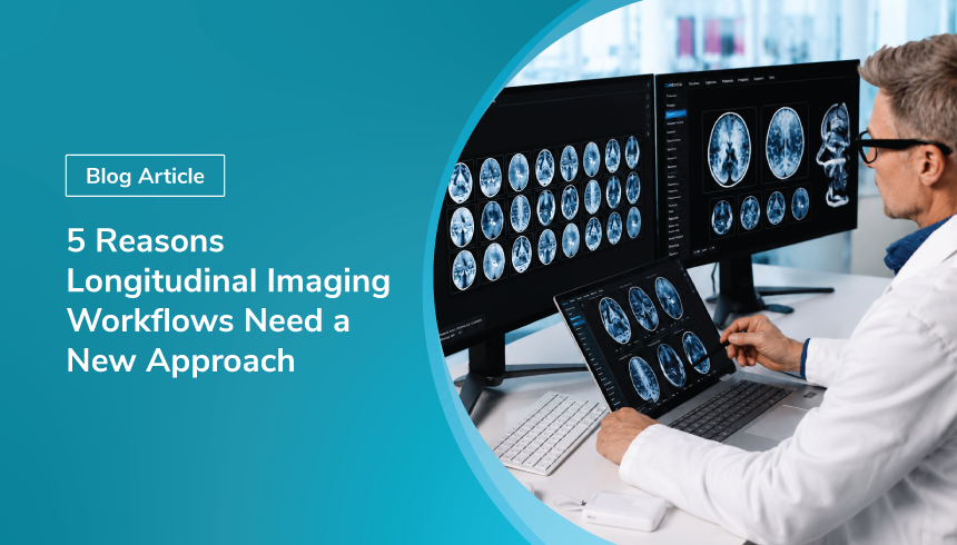
Clinical trials in neuro-oncology are becoming increasingly complex. Patients may undergo imaging over months or even years, with every new assessment depending on everything that came before it. Yet many imaging review workflows still treat each scan as an isolated event.
That was the focus of our recent webinar, "From Scattered Neuroimaging Time Points to One Table: How to Build the Full Subject Timeline Automatically," where Dr. Aly H. Abayazeed, Founder and CEO of QRad AI, joined Paulo Rodrigues, CTO and Co-founder of QMENTA, to discuss why longitudinal imaging requires a different operational approach.
Rather than reviewing individual scans, the conversation explored how imaging teams can build complete patient timelines that support more consistent reads, reduce operational burden, and improve oversight throughout a clinical trial.
Here are five of the biggest takeaways from the discussion.
1. Longitudinal Imaging Is About Patients, Not Individual Scans
Unlike cross-sectional studies, longitudinal clinical trials require radiologists to interpret every new examination within the context of a patient's complete imaging history.
Previous measurements, lesion annotations, response assessments, and earlier imaging visits all contribute to the interpretation of the current scan.
Dr. Abayazeed explained what’s most important:
"The ability to have this longitudinal view of a patient timeline when we're focusing on imaging and to be able to come up with accurate and valid decisions in real time."
Without that context immediately available, reviewers often spend valuable time reconstructing patient history instead of focusing on image interpretation.
The challenge isn't simply storing historical data; it's presenting that information in a way that supports confident clinical decision-making.
2. Consistency Is Often a Bigger Challenge Than Reader Availability
One of the discussion's most interesting observations was that neuro-oncology trials generally have access to experienced readers. The greater challenge is helping those readers make consistent decisions throughout long-running studies.
As patient histories grow, so does the amount of information reviewers must consider. The problem becomes even more pronounced when studies span several years, and reader turnover occurs.
As Dr. Abayazeed noted:
"The same reviewer might not be the same person that reads all the time points for the same patient. And that introduces a lot of variability."
When historical context is fragmented across multiple systems or reports, maintaining consistency becomes increasingly difficult regardless of who performs the review.
3. Legacy Workflows Weren't Designed for Longitudinal Studies
Many imaging review systems were originally built around individual imaging visits rather than complete patient journeys.
As a result, reviewers often need to leave their reading session to search for prior examinations, compare historical measurements, or manually reconstruct the patient's timeline before reaching a decision.
That extra effort grows with every follow-up visit, increasing cognitive workload while creating more opportunities for inconsistent assessments.
These inefficiencies frequently remain hidden until quality control activities or database lock, when resolving discrepancies becomes significantly more time-consuming.
4. Subject-Centric Workflows Reduce Operational Burden Across the Entire Study
A central theme throughout the webinar was shifting from scan-centric workflows to subject-centric ones.
Instead of asking readers to manually assemble historical information, the platform should automatically present the complete patient timeline from the moment the review begins.
During a live demonstration, QMENTA showed how reviewers can navigate seamlessly between baseline and follow-up examinations while keeping measurements, lesion annotations, response assessments, and prior imaging history in a single workspace.
This approach helps reduce manual navigation while giving readers immediate access to the clinical context needed to make informed decisions.
The benefits also extend beyond radiologists. Study managers must coordinate reader assignments, maintain continuity throughout long-running trials, support adjudication workflows, and ensure imaging data remains organized as new visits are added.
By automatically incorporating each new imaging time point into the patient's longitudinal record, imaging teams spend less time managing workflows and more time focusing on study execution.
5. Traceability Is Becoming Increasingly Important
As imaging studies become more complex, maintaining a complete record of imaging activities is no longer simply an administrative task.
Uploads, reader assignments, measurements, adjudications, exports, and administrative actions all contribute to the overall integrity of an imaging endpoint.
Rather than reconstructing these events retrospectively, QMENTA captures them automatically as part of the review process, creating a transparent audit trail throughout the study.
This level of traceability not only supports operational oversight but also helps study teams prepare for quality reviews and regulatory inspections with greater confidence.
Looking Ahead
One message stood out throughout the webinar: successful longitudinal imaging depends on connecting every imaging time point into a complete patient story—not treating each examination as an isolated event.
As neuro-oncology studies continue to become longer and more data-intensive, imaging workflows must evolve to provide reviewers with the full context they need while reducing the operational complexity faced by study teams.
As Dr. Abayazeed summarized:
"A system like [QMENTA] will likely help readers make better decisions, and better decisions usually mean a better chance of success for these trials."
That idea captures the central takeaway from the discussion. Better longitudinal imaging workflows aren't simply about reviewing scans more efficiently—they're about giving every stakeholder, from radiologists to study managers, the information they need to make better decisions throughout the life of a clinical trial.
Frequently Asked Questions
What are longitudinal imaging workflows?
Longitudinal imaging workflows are clinical trial imaging processes that connect multiple imaging time points for the same patient, allowing reviewers to assess each new scan in the context of the complete patient timeline.
Why are longitudinal imaging workflows important in neuro-oncology trials?
Longitudinal imaging workflows are important because neuro-oncology trials often require repeated imaging over months or years, with each assessment depending on previous measurements, lesion annotations, response assessments, and imaging history.
What is the difference between scan-centric and subject-centric imaging workflows?
Scan-centric workflows treat each imaging visit as a separate event, while subject-centric workflows organize imaging data around the patient journey, giving reviewers access to baseline, follow-up, measurements, annotations, and prior assessments in one workspace.
How can longitudinal imaging workflows reduce reader variability?
Longitudinal imaging workflows can reduce reader variability by making prior imaging history, measurements, lesion annotations, and response assessments easier to access, helping reviewers make more consistent decisions across time points.
Why is traceability important in clinical trial imaging?
Traceability is important because uploads, reader assignments, measurements, adjudications, exports, and administrative actions all contribute to the integrity of an imaging endpoint and support quality reviews and regulatory inspections.
Explore Longitudinal Imaging Workflows
Learn how QMENTA supports subject-centric imaging review, complete patient timelines, reader continuity, adjudication workflows, and traceable imaging data management across clinical trials.
Learn More